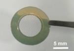
Extensive efforts have been devoted to develop new substrates for culture and differentiation of human induced pluripotent stem cells (hiPSCs) toward cardiac cell-based assays. A more exciting prospect is the construction of cardiac tissue for robust drug screening and cardiac tissue repairing. Here, we developed a patch method by electrospinning and crosslinking of monolayer gelatin nanofibers on a honeycomb frame made of poly(ethylene glycol) diacrylate (PEGDA). The monolayer of the nanofibrous structure can support cells with minimal exogenous contact and a maximal efficiency of cell–medium exchange whereas a single hiPSC colony can be uniformly formed in each of the honeycomb compartments. By modulating the treatment time of the ROCK inhibitor Y-27632, the shape of the hiPSC colony could be controlled from a flat layer to a hemisphere. Afterwards, the induction and differentiation of hiPSCs were achieved on the same patch, leading to a uniform cardiac layer with homogeneous contraction. This cardiac layer could then be used for extracellular recording with a commercial multi-electrode array, showing representative field potential waveforms of matured cardiac tissues with appropriate drug responses.

N’hésitez pas à consulter le communiqué de presse relatif à cet article : Des cellules souches pour reconstruire un coeur !
References:
Induction and differentiation of human induced pluripotent stem cells into functional cardiomyocytes on a compartmented monolayer of gelatin nanofibers
Yadong Tang, Li Liu, Junjun Li, Leqian Yu, Li Wang, Jian Shi and Yong Chen
Nanoscale, 2016, 8, 14530-14540
DOI : 10.1039/C6NR04545F
Induction and differentiation of human induced pluripotent stem cells into functional cardiomyocytes on a compartmented monolayer of gelatin nanofibers