2012
|
A New View of Electrochemistry at Highly Oriented Pyrolytic Graphite Article de journal Anisha N Patel; Manon Guille-Collignon; Michael A O'Connell; Wendy O Y Hung; Kim McKelvey; Julie V Macpherson; Patrick R Unwin Journal of the American Chemical Society, 134 (49), p. 20117-20130, 2012, (Times Cited: 157). @article{,
title = {A New View of Electrochemistry at Highly Oriented Pyrolytic Graphite},
author = {Anisha N Patel and Manon Guille-Collignon and Michael A O'Connell and Wendy O Y Hung and Kim McKelvey and Julie V Macpherson and Patrick R Unwin},
year = {2012},
date = {2012-01-01},
journal = {Journal of the American Chemical Society},
volume = {134},
number = {49},
pages = {20117-20130},
note = {Times Cited: 157},
keywords = {},
pubstate = {published},
tppubtype = {article}
}
|
Evaluation of the anti-oxidant properties of a SOD-mimic Mn-complex in activated macrophages Article de journal Anne-Sophie Bernard; Claire Giroud; Vincent H Y Ching; Anne Meunier; Vinita Ambike; Christian Amatore; Manon Guille-Collignon; Frederic Lemaitre; Clotilde Policar Dalton Transactions, 41 (21), p. 6399-6403, 2012, (Times Cited: 23). @article{,
title = {Evaluation of the anti-oxidant properties of a SOD-mimic Mn-complex in activated macrophages},
author = {Anne-Sophie Bernard and Claire Giroud and Vincent H Y Ching and Anne Meunier and Vinita Ambike and Christian Amatore and Manon Guille-Collignon and Frederic Lemaitre and Clotilde Policar},
year = {2012},
date = {2012-01-01},
journal = {Dalton Transactions},
volume = {41},
number = {21},
pages = {6399-6403},
note = {Times Cited: 23},
keywords = {},
pubstate = {published},
tppubtype = {article}
}
|
Indium Tin Oxide devices for amperometric detection of vesicular release by single cells Article de journal A Meunier; R Fulcrand; F Darchen; M Guille-Collignon; F Lemaître; C Amatore Biophysical Chemistry, 162 , p. 14–21, 2012. @article{Meunier:2012,
title = {Indium Tin Oxide devices for amperometric detection of vesicular release by single cells},
author = {A Meunier and R Fulcrand and F Darchen and M Guille-Collignon and F Lema\^{i}tre and C Amatore},
url = {https://www.scopus.com/inward/record.uri?eid=2-s2.0-84857369437&doi=10.1016%2fj.bpc.2011.12.002&partnerID=40&md5=47662a922db0ab7f5679e2054ed78ffb},
doi = {10.1016/j.bpc.2011.12.002},
year = {2012},
date = {2012-01-01},
journal = {Biophysical Chemistry},
volume = {162},
pages = {14--21},
abstract = {The microfabrication and successful testing of a series of three ITO (Indium Tin Oxide) microsystems for amperometric detection of cells exocytosis are reported. These microdevices have been optimized in order to simultaneously (i) enhance signal-to-noise ratios, as required electrochemical monitoring, by defining appropriate electrodes geometry and size, and (ii) provide surface conditions which allow cells to be cultured over during one or two days, through apposite deposition of a collagen film. The intrinsic electrochemical quality of the microdevices as well as the effect of different collagen treatments were assessed by investigating the voltammetric responses of two classical redox systems, Ru(NH 3) 6 3+/2 + and Fe(CN) 6 3-/4 -. This established that a moderate collagen treatment does not incur any significant alteration of voltammetric responses or degradation of the excellent signal-to-noise ratio. Among these three microdevices, the most versatile one involved a configuration in which the ITO microelectrodes were delimited by a microchannel coiled into a spiral. Though providing extremely good electrochemical responses this specific design allowed proper seeding and culture of cells permitting either single cell or cell cluster stimulation and analysis. © 2011 Elsevier B.V. All rights reserved.},
keywords = {},
pubstate = {published},
tppubtype = {article}
}
The microfabrication and successful testing of a series of three ITO (Indium Tin Oxide) microsystems for amperometric detection of cells exocytosis are reported. These microdevices have been optimized in order to simultaneously (i) enhance signal-to-noise ratios, as required electrochemical monitoring, by defining appropriate electrodes geometry and size, and (ii) provide surface conditions which allow cells to be cultured over during one or two days, through apposite deposition of a collagen film. The intrinsic electrochemical quality of the microdevices as well as the effect of different collagen treatments were assessed by investigating the voltammetric responses of two classical redox systems, Ru(NH 3) 6 3+/2 + and Fe(CN) 6 3-/4 -. This established that a moderate collagen treatment does not incur any significant alteration of voltammetric responses or degradation of the excellent signal-to-noise ratio. Among these three microdevices, the most versatile one involved a configuration in which the ITO microelectrodes were delimited by a microchannel coiled into a spiral. Though providing extremely good electrochemical responses this specific design allowed proper seeding and culture of cells permitting either single cell or cell cluster stimulation and analysis. © 2011 Elsevier B.V. All rights reserved. |
Nanoelectrodes for Determination of Reactive Oxygen and Nitrogen Species inside Murine Macrophages Article de journal Yixian Wang; Jean-Marc Noel; Jeyavel Velmurugan; Wojciech Nogala; Michael V Mirkin; Cong Lu; Manon Guille Collignon; Frederic Lemaitre; Christian Amatore Proceedings of the National Academy of Sciences of the United States of America, 109 (29), p. 11534-11539, 2012, ISSN: 0027-8424. @article{RN28b,
title = {Nanoelectrodes for Determination of Reactive Oxygen and Nitrogen Species inside Murine Macrophages},
author = {Yixian Wang and Jean-Marc Noel and Jeyavel Velmurugan and Wojciech Nogala and Michael V Mirkin and Cong Lu and Manon Guille Collignon and Frederic Lemaitre and Christian Amatore},
doi = {10.1073/pnas.1201552109},
issn = {0027-8424},
year = {2012},
date = {2012-01-01},
journal = {Proceedings of the National Academy of Sciences of the United States of America},
volume = {109},
number = {29},
pages = {11534-11539},
keywords = {},
pubstate = {published},
tppubtype = {article}
}
|
2011
|
Artificial synapses and oxidative stress Article de journal C Amatore; S Arbault; M Guille; F Lemaître Actualite Chimique, (348-349), p. 25–31, 2011. @article{Amatore:2011a,
title = {Artificial synapses and oxidative stress},
author = {C Amatore and S Arbault and M Guille and F Lema\^{i}tre},
url = {https://www.scopus.com/inward/record.uri?eid=2-s2.0-80052988658&partnerID=40&md5=d58fc6ddbe08657329d14270cacd4b93},
year = {2011},
date = {2011-01-01},
journal = {Actualite Chimique},
number = {348-349},
pages = {25--31},
abstract = {Free radical production in aerobic living beings is generally perceived only through a negative viewpoint since one focuses mostly on their deleterious effects. Yet, oxidative stress is an essential mechanism underlying many important functions in aerobic organisms including non specific immunedefenses or many regulations. This is mostly true for the primary species of oxidative stress, namely the superoxide anion and nitric oxide. However,any direct investigation of the production of these primary species was hampered up to the introduction by our group of the .artificial synapse. in this context. This article describes in first instance the principle of the method and justifies its high analytical performance. It focuses then onto its application to the investigation of two central mechanisms relying on oxidative stress: phagocytosis, active in macrophages, which provides non-specific means of fighting against microbial infections, and neurovascular coupling in brain, which allows our neurons to regulate their blood supply asa function of their activity and which is at the very basis of the current imaging techniques of brain activity.},
keywords = {},
pubstate = {published},
tppubtype = {article}
}
Free radical production in aerobic living beings is generally perceived only through a negative viewpoint since one focuses mostly on their deleterious effects. Yet, oxidative stress is an essential mechanism underlying many important functions in aerobic organisms including non specific immunedefenses or many regulations. This is mostly true for the primary species of oxidative stress, namely the superoxide anion and nitric oxide. However,any direct investigation of the production of these primary species was hampered up to the introduction by our group of the .artificial synapse. in this context. This article describes in first instance the principle of the method and justifies its high analytical performance. It focuses then onto its application to the investigation of two central mechanisms relying on oxidative stress: phagocytosis, active in macrophages, which provides non-specific means of fighting against microbial infections, and neurovascular coupling in brain, which allows our neurons to regulate their blood supply asa function of their activity and which is at the very basis of the current imaging techniques of brain activity. |
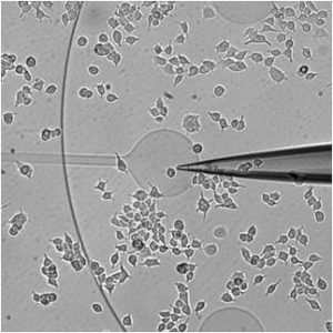
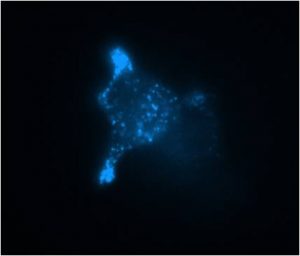
Coupling amperometry and total internal reflection fluorescence microscopy at ITO surfaces for monitoring exocytosis of single vesicles Article de journal A Meunier; O Jouannot; R Fulcrand; I Fanget; M Bretou; E Karatekin; S Arbault; M Guille; F Darchen; F Lemaître; C Amatore Angewandte Chemie - International Edition, 50 (22), p. 5081–5084, 2011. @article{Meunier:2011,
title = {Coupling amperometry and total internal reflection fluorescence microscopy at ITO surfaces for monitoring exocytosis of single vesicles},
author = {A Meunier and O Jouannot and R Fulcrand and I Fanget and M Bretou and E Karatekin and S Arbault and M Guille and F Darchen and F Lema\^{i}tre and C Amatore},
url = {https://www.scopus.com/inward/record.uri?eid=2-s2.0-79956075610&doi=10.1002%2fanie.201101148&partnerID=40&md5=3b903d6f37bf9d7fde5edb97402f8f0d},
doi = {10.1002/anie.201101148},
year = {2011},
date = {2011-01-01},
journal = {Angewandte Chemie - International Edition},
volume = {50},
number = {22},
pages = {5081--5084},
abstract = {More transparency in bioanalysis: A microdevice based on transparent indium tin oxide (ITO) electrodes allows simultaneous total internal reflection fluorescence microscopy and amperometric measurements. Use of the device in the coupled optical and electrochemical detection of single exocytotic events is demonstrated with enterochromaffin BON cells (see picture). Copyright © 2011 WILEY-VCH Verlag GmbH & Co. KGaA, Weinheim.},
keywords = {},
pubstate = {published},
tppubtype = {article}
}
More transparency in bioanalysis: A microdevice based on transparent indium tin oxide (ITO) electrodes allows simultaneous total internal reflection fluorescence microscopy and amperometric measurements. Use of the device in the coupled optical and electrochemical detection of single exocytotic events is demonstrated with enterochromaffin BON cells (see picture). Copyright © 2011 WILEY-VCH Verlag GmbH & Co. KGaA, Weinheim. |
2010
|
In situ electrochemical monitoring of reactive oxygen and nitrogen species released by single MG63 osteosarcoma cell submitted to a mechanical stress Article de journal Ren Hu; Manon Guille; Stephane Arbault; Chang Jian Lin; Christian Amatore Physical Chemistry Chemical Physics, 12 (34), p. 10048-10054, 2010, (Times Cited: 15). @article{,
title = {In situ electrochemical monitoring of reactive oxygen and nitrogen species released by single MG63 osteosarcoma cell submitted to a mechanical stress},
author = {Ren Hu and Manon Guille and Stephane Arbault and Chang Jian Lin and Christian Amatore},
year = {2010},
date = {2010-01-01},
journal = {Physical Chemistry Chemical Physics},
volume = {12},
number = {34},
pages = {10048-10054},
note = {Times Cited: 15},
keywords = {},
pubstate = {published},
tppubtype = {article}
}
|
Prediction of Local pH Variations during amperometric monitoring of vesicular exocytotic events at chromaffin cells Article de journal C Amatore; S Arbault; Y Bouret; M Guille; F Lemaître ChemPhysChem, 11 (13), p. 2931–2941, 2010. @article{Amatore:2010c,
title = {Prediction of Local pH Variations during amperometric monitoring of vesicular exocytotic events at chromaffin cells},
author = {C Amatore and S Arbault and Y Bouret and M Guille and F Lema\^{i}tre},
url = {https://www.scopus.com/inward/record.uri?eid=2-s2.0-77956858616&doi=10.1002%2fcphc.201000102&partnerID=40&md5=3e7bbeb9ad6306366c33ae3eebb2b086},
doi = {10.1002/cphc.201000102},
year = {2010},
date = {2010-01-01},
journal = {ChemPhysChem},
volume = {11},
number = {13},
pages = {2931--2941},
abstract = {Electrochemical monitoring of the exocytosis process is generally performed through amperometric oxidation of the electroactive messengers released by single living cells. Herein, we consider the vesicular release of catecholamines by chromaffin cells. Each exocytotic event is thus detected as a current spike whose morphology (intensity, duration, area, etc.) features the efficiency of the secretion process. However, the electrochemical oxidation of catechols produces quinone derivatives and protons. As a consequence, unless specific mechanisms may be adopted by a cell to regulate the pH near its membrane, the local pH between the cell membrane and the electrode necessarily drops within the electrode-cell cleft. Though this consequence of amperometric detection is generally ignored, it has been investigated in this work through simulation of the local pH drop created during the amperometric recording of a sequence of exocytotic events. This was performed based on frequencies and magnitudes of release detected at chromaffin cells. The corresponding acidification was shown to severely depend on the microelectrode radius. For usual 10 mm diameter carbon fiber electrodes, pH values below six were predicted to be reached within the electrode-cell cleft after monitoring a few current spikes. © 2010 Wiley-VCH Verlag GmbH& Co. KGaA, Weinheim.},
keywords = {},
pubstate = {published},
tppubtype = {article}
}
Electrochemical monitoring of the exocytosis process is generally performed through amperometric oxidation of the electroactive messengers released by single living cells. Herein, we consider the vesicular release of catecholamines by chromaffin cells. Each exocytotic event is thus detected as a current spike whose morphology (intensity, duration, area, etc.) features the efficiency of the secretion process. However, the electrochemical oxidation of catechols produces quinone derivatives and protons. As a consequence, unless specific mechanisms may be adopted by a cell to regulate the pH near its membrane, the local pH between the cell membrane and the electrode necessarily drops within the electrode-cell cleft. Though this consequence of amperometric detection is generally ignored, it has been investigated in this work through simulation of the local pH drop created during the amperometric recording of a sequence of exocytotic events. This was performed based on frequencies and magnitudes of release detected at chromaffin cells. The corresponding acidification was shown to severely depend on the microelectrode radius. For usual 10 mm diameter carbon fiber electrodes, pH values below six were predicted to be reached within the electrode-cell cleft after monitoring a few current spikes. © 2010 Wiley-VCH Verlag GmbH& Co. KGaA, Weinheim. |
Striking Inflammation from Both Sides: Manganese(II) Pentaazamacrocyclic SOD Mimics Act Also as Nitric Oxide Dismutases: A Single-Cell Study Article de journal Milos R Filipovic; Alaric C W Koh; Stephane Arbault; Vesna Niketic; Andrea Debus; Ulrike Schleicher; Christian Bogdan; Manon Guille; Frederic Lemaitre; Christian Amatore; Ivana Ivanovic-Burmazovic Angewandte Chemie-International Edition, 49 (25), p. 4228-4232, 2010, ISSN: 1433-7851. @article{RN23b,
title = {Striking Inflammation from Both Sides: Manganese(II) Pentaazamacrocyclic SOD Mimics Act Also as Nitric Oxide Dismutases: A Single-Cell Study},
author = {Milos R Filipovic and Alaric C W Koh and Stephane Arbault and Vesna Niketic and Andrea Debus and Ulrike Schleicher and Christian Bogdan and Manon Guille and Frederic Lemaitre and Christian Amatore and Ivana {Ivanovic-Burmazovic}},
doi = {10.1002/anie.200905936},
issn = {1433-7851},
year = {2010},
date = {2010-01-01},
journal = {Angewandte Chemie-International Edition},
volume = {49},
number = {25},
pages = {4228-4232},
keywords = {},
pubstate = {published},
tppubtype = {article}
}
|
2009
|
Invariance of exocytotic events detected by amperometry as a function of the carbon fiber microelectrode diameter Article de journal C Amatore; S Arbault; Y Bouret; M Guille; F Lemaître; Y Verchier Analytical Chemistry, 81 (8), p. 3087–3093, 2009. @article{Amatore:2009e,
title = {Invariance of exocytotic events detected by amperometry as a function of the carbon fiber microelectrode diameter},
author = {C Amatore and S Arbault and Y Bouret and M Guille and F Lema\^{i}tre and Y Verchier},
url = {https://www.scopus.com/inward/record.uri?eid=2-s2.0-65249125788&doi=10.1021%2fac900059s&partnerID=40&md5=3a9131b641fb9496ef04b7576ed24617},
doi = {10.1021/ac900059s},
year = {2009},
date = {2009-01-01},
journal = {Analytical Chemistry},
volume = {81},
number = {8},
pages = {3087--3093},
abstract = {Etched carbon fiber microelectrodes of different radii have been used for amperometric measurements of single exocytotic events occurring at adrenal chromaffin cells. Frequency, kinetic, and quantitative information on exo-cytosis provided by amperometric spikes were analyzed as a function of the surface area of the microelectrodes. Interestingly, the percentage of spikes with foot (as well as their own characteristics), a category revealing the existence of sufficient long-lasting fusion pores, was found to be constant whatever the microelectrode diameter was, whereas the probability of overlapping spikes decreased with the electrode size. This confirmed that the prespike foot could not feature accidental superimposition of separated events occurring at different places. Moreover, the features of amperometric spikes investigated here (charge, intensity and kinetics) were found constant for all microelectrode diameters. This demonstrated that the electrochemical measurement does not introduce significant bias onto the kinetics and thermodynamics of release during individual exocytotic events. All in all, this work evidences that information on exocytosis amperometri-cally recorded with the usual 7 μm diameter carbon fiber electrodes is biologically relevant, although the frequent overlap between spikes requires a censorship of the data during the analytical treatment. © 2009 American Chemical Society.},
keywords = {},
pubstate = {published},
tppubtype = {article}
}
Etched carbon fiber microelectrodes of different radii have been used for amperometric measurements of single exocytotic events occurring at adrenal chromaffin cells. Frequency, kinetic, and quantitative information on exo-cytosis provided by amperometric spikes were analyzed as a function of the surface area of the microelectrodes. Interestingly, the percentage of spikes with foot (as well as their own characteristics), a category revealing the existence of sufficient long-lasting fusion pores, was found to be constant whatever the microelectrode diameter was, whereas the probability of overlapping spikes decreased with the electrode size. This confirmed that the prespike foot could not feature accidental superimposition of separated events occurring at different places. Moreover, the features of amperometric spikes investigated here (charge, intensity and kinetics) were found constant for all microelectrode diameters. This demonstrated that the electrochemical measurement does not introduce significant bias onto the kinetics and thermodynamics of release during individual exocytotic events. All in all, this work evidences that information on exocytosis amperometri-cally recorded with the usual 7 μm diameter carbon fiber electrodes is biologically relevant, although the frequent overlap between spikes requires a censorship of the data during the analytical treatment. © 2009 American Chemical Society. |
Quantitative investigations of amperometric spike feet suggest different controlling factors of the fusion pore in exocytosis at chromaffin cells Article de journal Christian Amatore; Stephane Arbault; Imelda Bonifas; Manon Guille Biophysical Chemistry, 143 (3), p. 124-131, 2009, (Times Cited: 25). @article{,
title = {Quantitative investigations of amperometric spike feet suggest different controlling factors of the fusion pore in exocytosis at chromaffin cells},
author = {Christian Amatore and Stephane Arbault and Imelda Bonifas and Manon Guille},
year = {2009},
date = {2009-01-01},
journal = {Biophysical Chemistry},
volume = {143},
number = {3},
pages = {124-131},
note = {Times Cited: 25},
keywords = {},
pubstate = {published},
tppubtype = {article}
}
|
2008
|
Electrochemical monitoring of single cell secretion: Vesicular exocytosis and oxidative stress Article de journal C Amatore; S Arbault; M Guille; F Lemaître Chemical Reviews, 108 (7), p. 2585–2621, 2008. @article{Amatore:2008a,
title = {Electrochemical monitoring of single cell secretion: Vesicular exocytosis and oxidative stress},
author = {C Amatore and S Arbault and M Guille and F Lema\^{i}tre},
url = {https://www.scopus.com/inward/record.uri?eid=2-s2.0-49049112285&doi=10.1021%2fcr068062g&partnerID=40&md5=ae6a20b01b9ae57f75d37389b0ebe3ec},
doi = {10.1021/cr068062g},
year = {2008},
date = {2008-01-01},
journal = {Chemical Reviews},
volume = {108},
number = {7},
pages = {2585--2621},
abstract = {Several important contributions of electroanalytical techniques over the past 20 years for investigating three major biological processes at the single cell level: vesicular exocytosis, oxidative stress, and nitric oxide metabolism in brain have been reported. It is evident that molecular electrochemistry at microelectrodes enhances the understanding of central processes of cellular biology including cellular metabolism either at a single cell stage or in living tissues. Since cells have highly variable metabolism even among single genetic lines, studies performed at the single cell level allow delineating precisely the extent and limits of these variabilities.},
keywords = {},
pubstate = {published},
tppubtype = {article}
}
Several important contributions of electroanalytical techniques over the past 20 years for investigating three major biological processes at the single cell level: vesicular exocytosis, oxidative stress, and nitric oxide metabolism in brain have been reported. It is evident that molecular electrochemistry at microelectrodes enhances the understanding of central processes of cellular biology including cellular metabolism either at a single cell stage or in living tissues. Since cells have highly variable metabolism even among single genetic lines, studies performed at the single cell level allow delineating precisely the extent and limits of these variabilities. |
2007
|
Relationship between amperometric pre-spike feet and secretion granule composition in Chromaffin cells: An overview Article de journal C Amatore; S Arbault; I Bonifas; M Guille; F Lemaître; Y Verchier Biophysical Chemistry, 129 (2-3), p. 181–189, 2007. @article{Amatore:2007n,
title = {Relationship between amperometric pre-spike feet and secretion granule composition in Chromaffin cells: An overview},
author = {C Amatore and S Arbault and I Bonifas and M Guille and F Lema\^{i}tre and Y Verchier},
url = {https://www.scopus.com/inward/record.uri?eid=2-s2.0-34547560678&doi=10.1016%2fj.bpc.2007.05.018&partnerID=40&md5=c92376d4195c4dfec6edfd9e5eb98173},
doi = {10.1016/j.bpc.2007.05.018},
year = {2007},
date = {2007-01-01},
journal = {Biophysical Chemistry},
volume = {129},
number = {2-3},
pages = {181--189},
abstract = {Amperometry is a simple and powerful technique to study exocytosis at the single cell level. By positioning and polarizing (at an appropriate potential at which the molecules released by the cell can be oxidized) a carbon fiber microelectrode at the top of the cell, each exocytotic event is detected as an amperometric spike. More particularly, a portion of these spikes has previously been shown to present a foot, i.e. a small pedestal of current that precedes the spike itself. Among the important number of works dealing with the monitoring of exocytosis by amperometry under different conditions, only a few studies focus on amperometric spikes with a foot. In this work, by coupling our previous and recent experiments on chromaffin cells (that release catecholamines after stimulation) with literature data, we bring more light on what an amperometric foot and particularly its features, represents. © 2007.},
keywords = {},
pubstate = {published},
tppubtype = {article}
}
Amperometry is a simple and powerful technique to study exocytosis at the single cell level. By positioning and polarizing (at an appropriate potential at which the molecules released by the cell can be oxidized) a carbon fiber microelectrode at the top of the cell, each exocytotic event is detected as an amperometric spike. More particularly, a portion of these spikes has previously been shown to present a foot, i.e. a small pedestal of current that precedes the spike itself. Among the important number of works dealing with the monitoring of exocytosis by amperometry under different conditions, only a few studies focus on amperometric spikes with a foot. In this work, by coupling our previous and recent experiments on chromaffin cells (that release catecholamines after stimulation) with literature data, we bring more light on what an amperometric foot and particularly its features, represents. © 2007. |
The Nature and Efficiency of Neurotransmitter Exocytosis Also Depend on Physicochemical Parameters Article de journal Christian Amatore; Stephane Arbault; Manon Guille; Frederic Lemaitre Chemphyschem, 8 (11), p. 1597-1605, 2007, ISSN: 1439-4235. @article{RN21b,
title = {The Nature and Efficiency of Neurotransmitter Exocytosis Also Depend on Physicochemical Parameters},
author = {Christian Amatore and Stephane Arbault and Manon Guille and Frederic Lemaitre},
doi = {10.1002/cphc.200700225},
issn = {1439-4235},
year = {2007},
date = {2007-01-01},
journal = {Chemphyschem},
volume = {8},
number = {11},
pages = {1597-1605},
keywords = {},
pubstate = {published},
tppubtype = {article}
}
|
2006
|
Assessment of the electrochemical behavior of two-dimensional networks of single-walled carbon nanotubes Article de journal Neil R Wilson; Manon Guille; Ioana Dumitrescu; Virginia R Fernandez; Nicola C Rudd; Cara G Williams; Patrick R Unwin; Julie V Macpherson Analytical Chemistry, 78 (19), p. 7006-7015, 2006, (Times Cited: 28). @article{,
title = {Assessment of the electrochemical behavior of two-dimensional networks of single-walled carbon nanotubes},
author = {Neil R Wilson and Manon Guille and Ioana Dumitrescu and Virginia R Fernandez and Nicola C Rudd and Cara G Williams and Patrick R Unwin and Julie V Macpherson},
year = {2006},
date = {2006-01-01},
journal = {Analytical Chemistry},
volume = {78},
number = {19},
pages = {7006-7015},
note = {Times Cited: 28},
keywords = {},
pubstate = {published},
tppubtype = {article}
}
|


