For proteins in solvent mixtures, the relative abundances of each solvent in their solvation shell have a critical impact on their properties. Preferential solvation of a series of proteins in water–glycerol mixtures is studied here over a broad range of solvent compositions via classical molecular dynamics simulations. Our simulation results reveal that the differences between shell and bulk compositions exhibit dramatic changes with solvent composition, temperature, and protein nature. In contrast with the simple and widely used picture where glycerol is completely excluded from the protein interface, we show that for aqueous solutions with less than 50% glycerol in volume, protein solvation shells have approximately the same composition as the bulk solvent and proteins are in direct contact with glycerol. We further demonstrate that at high glycerol concentration, glycerol depletion from the solvation shell is due to an entropic factor arising from the reduced accessibility of bulky glycerol molecules in protein cavities. The resulting molecular picture is important to understand protein activity and cryopreservation in mixed aqueous solvents.
References:
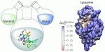
Protein Preferential Solvation in Water:Glycerol Mixtures
Nicolas Chéron, Margaux Naepels, Eva Pluhařová and Damien Laage
J. Phys. Chem. B 2020, 124, 8, 1424-1437
doi: 10.1021/acs.jpcb.9b11190