Kinetic Redox Shotgun Proteomics Reveals Specific Lipopolysaccharide Effects on Intestinal Epithelial Cells, Mitigated by a Mn Superoxide Dismutase Mimic
Martha Zoumpoulaki 1-3, Giovanni Chiappetta 2, Jean Bouvet 1, Namita-Raju John 1, Gabrielle Schanne 1,3, Pauline Gehan 1, Samuel Diebolt 2, Shakir Shakir 2, Elodie Quévrain 1,3, Emilie Mathieu 1, Sylvie Demignot 3,4, Philippe Seksik 3, Nicolas Delsuc 1, Joelle Vinh 2,* and Clotilde Policar 1,*
1 Laboratoire Chimie Physique et chimie du vivant—CPCV, UMR8228, Département de Chimie, Ecole Normale Supérieure, PSL University, Sorbonne Université, CNRS, 75005 Paris, France
2 Laboratoire de Spectrométrie de Masse Biologique et Protéomique, SMBP, PdC UMR8249, ESPCI Paris, PSL University, CNRS, 75005 Paris, France
3 Gastroenterology Department, Sorbonne Université, INSERM, Centre de Recherche Saint-Antoine, CRSA, Paris Center for Microbiome Medicine (PaCeMM) FHU, AP-HP, Saint-Antoine Hospital, Paris, France
4 EPHE, PSL University, 75014 Paris, France
* Correspondance e-mail : joelle.vinh@espci.psl.eu (0000-0001-7184-2668) & clotilde.policar@ens.psl.eu (0000-0003-0255-1650)
Angew. Chem. Int. Ed. 2025, e202422644 doi.org/10.1002/anie.202422644
Context: The overproduction of reactive oxygen species (ROS) and the dysregulation of antioxidant superoxide dismutases (SOD) such as the Mn-based SOD2 contribute to chronic inflammation such as generated in inflammatory bowel diseases (IBD).
At the Ecole Normale Supérieure, in the METHROX research group (https://ens-bic.fr/), we are developing both low molecular-weight Mn-complexes capable of complementing SOD2,[1] and cellular models to screen them.[1,2] In particular, bacterial lipopolysaccharide (LPS) has been shown to induce a very intense inflammation on a cellular model of intestinal epithelial cells (HT29-MD2) built in the Centre de Recherche de l’Hôpital Saint Antoine. HT29-MD2 responds strongly to LPS that triggers an intense inflammation (measured by interleukin 8 IL8 and cyclooxygenase 2 COX2).[3]
The SOD mimics Mn1 (see figure below) was shown to be able to limit the LPS-induced inflammation as measured with the quantification these inflammatory markers when co-incubated with LPS.[1,4,5] Interestingly, LPS challenge leads to the overexpression of SOD2 in an active form. This was interpreted as an indirect proof that LPS was inducing a superoxide-based oxidative stress. The overexpression of SOD2 can be considered as a cellular feedback to fight oxidative stress.
Coincubation of LPS with Mn1 was shown to attenuate the SOD2 overexpression,[1,4,5] consistent with the facts that (a) LPS-induced inflammation is mediated by oxidative stress and probably by superoxide, and (b) Mn1 is able to complement for SOD2.
Present work: We wanted to study both the effect of LPS and LPS-Mn1 co-incubation, without any a priori, by looking at the proteome. We also wanted to quantify the effect on the redox cellular status using the cysteine redoxome as a reversible marker for oxidative stress at different time points (15 min to 6h time scales).
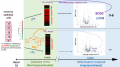
A kinetic redox shotgun proteomic strategy (labelled OcSILAC for Oxidized cysteine Stable Isotope Labelling by Amino acids in Cell culture), set up in the Spectrométrie de masse biologique (ESPCI) laboratory, was used to explore the LPS effects on HT29-MD2 cells and those of Mn1 coincubation. A cellular fractionation enabled us to maximize the protein coverage and shed special light on the mitochondrial fraction.
Interestingly, LPS induced transient Cys oxidation at early times (15-30 min). Cells co-incubated with Mn1 attenuated this early cysteine oxidation, highlighting Mn1 antioxidant cellular properties. We validated these results by fluorescence quantification of Cys reversible oxidation in cell lysates (on gel).
Over time, cysteine oxidation of LPS treated cells was counteracted by an overexpression of antioxidant proteins (SOD1, NQO1) and a late (6h) preponderant increased in SOD2 level. Mn1, when co-incubated with LPS, attenuated the level of most LPS-modified proteins, mainly proteins involved in the inflammatory response.
The effects we observed, particularly on oxidative stress, are moderate: this was anticipated as the intestinal epithelial HT29-MD2 cell-line is not designed to produce high levels of ROS, as could have done macrophages. However, the relevance of this cell line to reductionist models of IBD makes them interesting to study. The powerful OcSILAC approach allowed us to monitor these moderate but biologically relevant effects. This work shows an unambiguous direct link between oxidative stress and inflammation: they are not only associated but oxidative stress precedes inflammation. It indicates also the SOD mimics Mn1 is an effective antioxidant and anti-inflammatory agent to be considered in the treatment of IBD, as well as a useful tool for exploring the interconnection between oxidative stress and inflammation.
