Living systems, such as cells, are characterized by a precise spatio-temporal distribution of their components. In order to understand the mechanisms that govern live cell functions, scientists sometimes build simplified models of cells, commonly known as synthetic cells or artificial cells. These systems, with a perfectly well-known composition and on which one can act at will, make it possible to study a variety of phenomena and understand certain essential elements. However, to date, most synthetic cells are globally isotropic objects, with no top or bottom, no left or right, no head or feet – a far cry from the multi-component, polarized structures of living cells.
We therefore started with one of the most widely-used cellular models, the giant unilamellar vesicle (GUV), and developed a method for not only polarizing it, but also controlling the number of its poles and positioning them in a user-defined manner. A GUV is a closed lipid bilayer separating an internal aqueous medium from another outer aqueous medium. Generally spherical in shape when unconstrainted, its size varies from a few micrometers to a few hundred micrometers, making it comparable to cellular dimensions. For this work, we used membranes composed of lipid mixtures leading to the coexistence of two phases: an liquid-ordered phase (Lo) and a liquid-disordered phase (Ld). If the Lo phase dominates, Ld domains are formed within the continuous Lo phase, and vice versa. This well-known phenomenon is often used to mimic domain formation in biological membranes. However, these domains are mobile, impossible to position, and tend to merge to minimize the energy of the contact line between the two phases. This results in a single, randomly positioned domain composed of the minority phase.
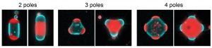
Figure 1. By confining giant unilamellar vesicles (GUVs) in different geometries, it is possible to position lipid domains deterministically, enabling the creation of cellular models with 2, 3 or 4 poles. In these images, the liquid-disordered (Ld) phase appears in red, the liquid-ordered (Lo) phase in blue.
To guide the organization and localization of these domains, we confined the GUVs in order to deform them and generate, within each GUV, some free curved zones so as to promote the fusion of minority-phase domains by minimizing line energy. We first confined the GUVs in microchannels and showed that the minority-phase domains accumulate systematically at the free curved ends and merge there to form a single domain at each extremity, thus creating a GUV with two “poles” (Figure 1, left). Interestingly, it’s not the chemical nature of the lipid that determines its position, but rather the fraction of its phase. It is thus possible to create Ld or Lo poles, yet having very different lipid compositions, simply by adjusting the respective fraction of the two phases. Using a variety of confinement geometries, including pillar arrays, we were finally able to create GUVs with 3 or 4 poles (Figure 1, middle and right) positioned in a deterministic manner.
This work shows for the first time how to control the number and position of poles, in the form of lipid domains, within GUVs used as simplified cell models. By coupling the position of these domains with other components (DNA, proteins), this method will make it possible to control or study, within model systems, the spatio-temporal organization of key elements involved in a variety of processes such as cytoskeletal dynamics, cell division, regulatory processes or compartmentalization. This work also highlights the fundamental role of line energy in self-organization phenomena within biological or reconstituted membranes.
Reference : K. Nakazawa, A. Lévrier, S. Rudiuk, A. Yamada, M. Morel, D. Baigl.* Controlled Lipid Domain Positioning and Polarization in Confined Minimal Cell Models. Angew. Chem. Int. Ed. 2025, e202419529
Link (open access) : https://doi.org/10.1002/anie.202419529
Contact : Damien Baigl (damien.baigl@ens.psl.eu)